SV inspector view
The SV inspector is a combined variant table and whole-genome circular view for triaging structural variant calls.
For an end-to-end walkthrough using real cancer sequencing data, see the C-GIAB tutorial.
Launch it from the main menu bar:
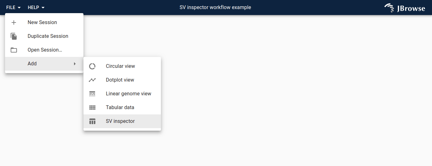
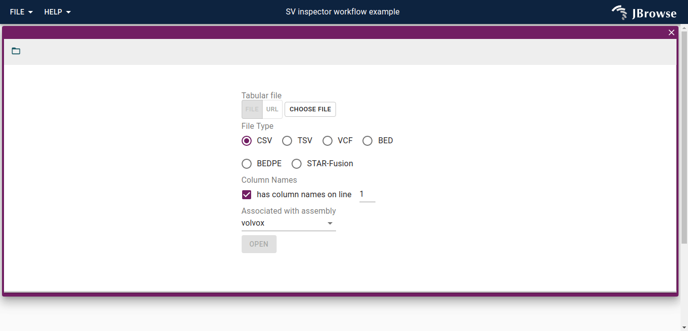
An import form will appear asking for your SV data.
The following formats are supported:
- CSV, TSV
- VCF or VCF.gz (plain text VCF, or (b)gzipped VCF)
- BED, BEDPE
- STAR-fusion result file
Sources of data for SV inspector
The SV inspector is designed for viewing <TRA> and breakend type entries.
Compatible variant callers:
Short read based:
- Manta
- Delly
- Lumpy
Long read based
- pbsv
- Sniffles
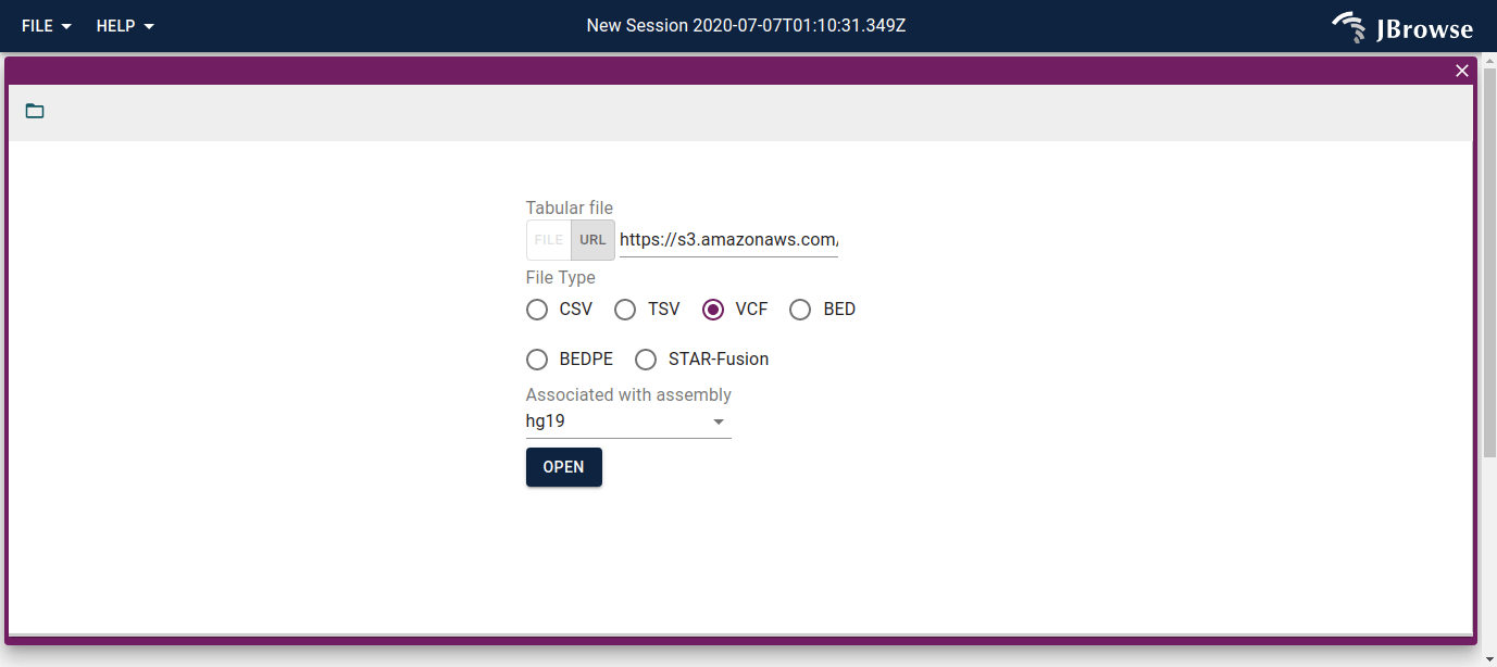
Example workflow
As an example, load this VCF of translocation events called from the SKBR3 breast cancer cell line (published data). Paste the URL into the import form and select hg19:
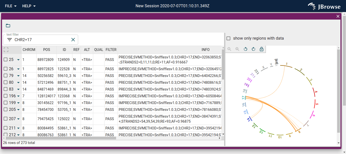
SV inspector results
The loaded file appears as a searchable table with each row representing one variant, alongside a whole-genome circular overview.
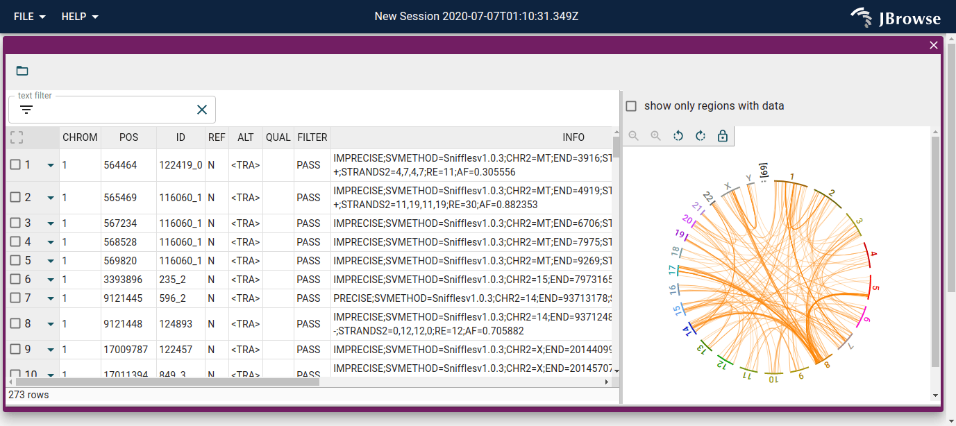
Table filters are reflected in the circular view.
Launching breakpoint split view
Click a feature in the circular view, or the triangle dropdown on any table row, to open the breakpoint split view for that variant.
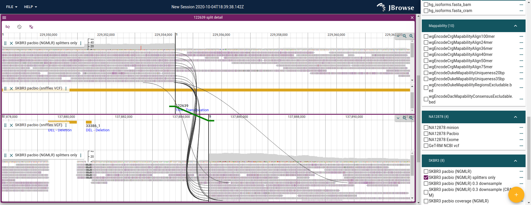
Loading alignment tracks
The breakpoint split view opens with empty top and bottom views. Add alignment tracks to both views using their track selectors (the tracks button in each view header). Read arcs and splines connecting supporting reads then appear automatically.